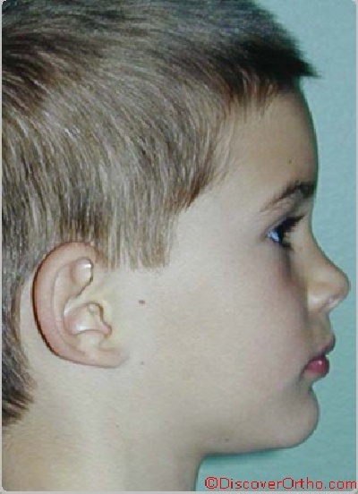


Facial Exam
Profile Analysis
Studying the patient’s profile directly or from a photograph will enable you to assess the facial convexity and give valuable information regarding the position of the facial structures in the antero-posterior plane.
It is essential to evaluate the profile when the patient is young because antero-posterior problems will usually not improve during active growth. This does not mean that all problems should be treated when they are detected, but parents should be informed of the malocclusion as soon as possible. Some treatment modalities will be better treated when the patient is young while others can be best corrected in the late mixed dentition or early adult dentition.
It is very important for you to understand the facial characteristics of your patients and their potential implications to start treatment at the best possible time. The treatment modality chosen must also respect the principles of facial harmony and not only the dento-alveolar relationships. Keep in mind that patients seek orthodontic care mainly to improve their facial esthetics. Orthodontic intervention must have a positive effect on facial balance. In order to improve or maintain facial esthetics, you must carefully assess the facial features of the patient and the benefits or limitations of orthodontic therapy.
Simple profile determination
We are going to use the patient's profile photograph to illustrate this analysis.
Horizontal reference line
First determine "soft tissue" Orbitale and trace a horizontal line through this point. This line is known as the "Frankfort horizontal".
Vertical reference line
Identify Glabella (Gl) and draw a vertical line from "Frankfort" going through Glabella. This reference line will allow us to estimate nasal projection and the relative position of the upper and lower lips and chin point.
Relative position of the maxilla and mandible
We need a reference point for each jaw to assess their relative position.
Maxillary reference point
The Subnasale point (SN) is located about 5mm under the base of the nose. It corresponds to the A point in the cephalometric analysis. If the Subnasale is located on or slightly in front of the reference line, and the lip is well developed, the maxilla is probably well positioned. If the Subnasale is located behind the reference line, the maxilla may be retrusive.
Mandibular reference point
The projection of the chin is referenced by the Pogonion soft, which is located on the most anterior point of the chin. It is normal for a growing child to have this point behind the reference line. If the point is positioned in front of the line, investigate the possibility of a Cl III or pseudo Cl III malocclusion.
If the Pogonion is severely behind the reference line, suspect mandibular retrognatism.
Profile convexity
The angle formed by the lines Glabella/Subnasale and Pogonion/Subnasale will allow the clinician to estimate the facial convexity. Relating this finding to the relative position of the maxilla and mandible to the reference line will help determine the etiology of the potential skeletal dysplasia.
If a patient presents a straight or slightly concave profile at the age of 8 years, further investigation (with cephalometry) may show that a skeletal Cl III is developing.
The patients showing severe profile convexity (of skeletal origin) should also be carefully monitored because a severe skeletal malocclusion may be present but the dental occlusal relationship appear fairly normal. In other words, the skeletal discrepancy is well camouflaged by favorable dento-alveolar compensations.
The profile analysis is only a qualitative tool and does not possess the accuracy of a cephalometric analysis. However, it will help you during the patient selection process.